Description of an industrial invention by title
Realization of antibacterial nanometric coating with controlled release of bactericidal agent
(Patent pending, No. WO 2013/021409 A8)
SUMMARY
The present invention relates to the realization of hydrophilic coatings with antibacterial properties of controlled, uniform and long-lasting release of Ag + ions in wet environments. "In vitro" has been demonstrated that coatings deposited via plasma with a structure similar to polyethylene oxide (PEO-like) polymers discourage protein adsorption and cell adhesion; Similar coatings, when they contain silver clusters, also have a distinct bactericidal effect, for example against P.aeuriginosa and S.epidermis bacteria.
It is based on the study of PE-CVD processes (Plasma Enhanced Chemical Vapor Deposition) for the deposition of organic, inorganic or nano-composite thin films (20 nanometers - 1 micron). By varying the plasma feed gas and the deposition conditions it is possible to obtain thin films with continuously variable composition with the deposition conditions, with absolutely unique characteristics, among which organic coatings with "non-fouling" resistance characteristics, ie, all adhesion and contamination with organic and biological substances in an aqueous medium (with "monomers" such as ethylene glycols). By coupling a sputtering process to a conventional PE-CVD process, it is possible to produce nanocomposite coatings, consisting, that is, of nanometric clusters (metallic, ceramic or polymeric) included in thin film.
STATE OF THE TECHNIQUE
The plasma deposition techniques of films with repellent properties for biological material are known. On the surface of a material that has affinity for biological components (proteins, cells, bacteria, fungi, algae), once it is placed in a non-sterile environment, it is formed a biofilm (fouling) of variable composition that can promote adhesion and growth of bacteria and spores. The presence of biomolecules can alter the performance of the device (sensor, filter, reactor, prosthesis) with which the material is manufactured. For biomedical devices such as vascular prostheses and catheters, fouling can degenerate into bacterial colonization, thus into infections, degradation of materials and removal of infected prostheses, with even serious consequences on patients.
The possibility of giving the film antibacterial characteristics has also been studied, through the use of fine dispersions, in globules of about 2-4 nm, of silver, an antibacterial known since ancient times, included in the matrix of a non-fouling film. , during a single deposition process. The antibacterial efficacy (bacterial population decrease of 6 orders of magnitude) was tested on lines of bacteria Staphilococcus Aureus and Pseudomonas Aeruginosa.
The release of the silver ions takes place, however, in an uncontrolled and rapidly decreasing manner, depending on the concentration of the globules in the coating, on the nature and thickness of the same and on the chemical-physical properties of the medium in which the device is placed.
The research activity of MEDIPLASMA together with the associated PLASMA SOLUTION (spin-ff of the University of Bari) and of UNIBA in this sector has allowed to develop a controlled, uniform and long-lasting, non-cytotoxic release device, which constitutes the essence of this patent, whose title is 75% for MEDIPLASMA and 25% for the University of Bari.
SCIENTIFIC FOUNDATION "POTITO" - UNIVERSITY OF MOLISE
The following are the summaries of three studies carried out by the POTITO scientific foundation on the chemical-physical, microbiochemical and immunological properties of the antibacterial coating during international patenting. Full studies are available at the request of interested parties.
Chemical-physical characterization study and modalities of the release of silver ions from nano-structured chemical plasmo coating, deposited on non-commercial samples of intraocular lentils (Prof. Luigi Ambrosone).
The study was carried out on non-commercial samples of intraocular lentils, set up at the Plasma Solution Company (spin off of the University of Bari) and covered with nanometric coatings with controlled release of silver ions, for which a request for national and international patent.
Several model systems have been created to be used as improvement, or substitutive, of vision devices (intraocular lenses, contact lenses). In this case, polyethylentrephthalate (PET) squares were coated, about 1 cm per side, with a nanomolecular Ag layer, with coating thickness in the range 10 - 100 nm.
By appropriately selecting the parameters of the "plasmo-chemical" process, both the quantity of embedded silver and the degree of cross-linking of the organic phase can be controlled, in which the inorganic (silver) is finely dispersed.
A first type of experiments showed that Ag-free PET immersed in 100 ml of a physiological solution, at a temperature of 37 ° C after about two months, did not release in solution Ag (in any form); the sensitivity of the device was 10 ppb (parts per billion).
The antibacterial effect of silver, therefore, can be modulated by varying the characteristics of the deposited film; therefore, three different types of samples were prepared. Their characteristics are reported in Tab. 1. These are three types of samples which essentially consist of Ag film not covered by any other Ag film and film covered with a further polyethylene oxide (PEO) layer at different percentages.
Tab.1 Different types of samples prepared with the APS technique on squares of 1 cm side of PET
TYPE I
Film Ag / PEO-like (Ag 5%) absence of protective film
TYPE II
Film Ag / PEO-like (Ag (5%) bfilm protective (PEO 70%)
TYPE III
Film Ag / PEO-like (Ag 5%) protective film (30% PEO)
Two types of analyzes were carried out: morphological / structural and chemical-physical. The former were conducted directly on solid samples using Scanning Electron Microscopy (SEM) and X-ray spectroscopy (XRD); the latter were carried out on the liquid solution in which the samples were immersed to evaluate their release properties.
Part One - Morphological measurements
- SEM analysis
The analyzes were performed with a powerful tool produced by Zeiss.
- X-ray diffraction (XRD)
On the microscope there is a detector (Oxford Instruments) for EDS analysis, Energy Dispersive X-ray. The identification of the peaks obtained was carried out thanks to comparisons with spectra present in the extensive software database.
Sample preparation
The samples were prepared by covering, with the plasmas technique, the PoliEtileneTerephthalate (PET) specimens of square shape with 1cm side. The final properties of the prepared material depend both on the quantity of Silver and other compounds used for the covering of the specimens, and on the modalities with which they are covered. Therefore, three types of specimens with different surface characteristics were studied.
1) Type I, marked by red color, built by covering the specimen with a film of Silver / Polyethylene oxide (PEO) at 5% Ag. The film is left free or without any additional coverage.
2) Type II, marked with a blue color, built by covering the specimen with a film of Silver / Polyethylene oxide (PEO) at 5% Ag. The film is further coated with a further 70% PEO film.
3) Type III, marked by the color black, built by covering the specimen with a film of Silver / Polyethylene oxide (PEO) at 5% of Ag. The film is further coated with a further 30% PEO film.
If you compare the results of the spectra for the three types of samples, you immediately notice how the samples with the further coating of PEO exhibit a lower percentage of Ag on the surface. This result can be explained bearing in mind that the penetration capacity of the electrons in the SEM is always the same in the three samples, but since the type I specimen does not have the covering PEO layer, the concentration of Ag atoms results in more high. In other words, the amount of silver contained in the first layer is referred to a volume approximately twice as large, corresponding to the set of two superimposed layers.
Part two - Release measures
Experimental method for the determination of silver in aqueous solution
The silver released from the samples at different times was determined by atomic absorption spectrophotometry. According to this method, silver is determined by direct injection of a sample into the graphite furnace of an atomic absorption spectrophotometer.
Kinetics of release of Ag in solution at pH 7.2 Temperature 37.0 ° C.
The three sample types behave differently, at the same observation time. In absolute terms, type III releases less Ag than do Type I and II. Type I seems to reach a plateau around 10 days, then re-grows at different speeds. In any case, it releases the highest concentration of Ag. For Type II samples, the release curve is similar to Type I, although the Ag concentration is slightly lower. Type III samples exhibit a completely different release curve, continuously increasing until reaching a plateau.
Type I samples are characterized by the absence of the protective film so that the Ag layer is in direct contact with the solution. In this case, the water molecules can freely enter the film, inflating it (swelling) and favoring the diffusion of the Ag towards the bulk solution. It is good to specify that silver can either diffuse as Ag or transform into ion (Ag +) generating a colloidal silver suspension. This interpretation also explains the ascent of the red curve. In fact, water molecules must spread inside the film before Ag's atoms can release; when the concentration of water inside the layer is sufficiently high, the concentration of Ag increases, which is "detached" from the film.
Samples II and III consist of a same Ag layer (5%) and protected with PEO film at different percentage concentrations. The difference in behavior shown in the graph, therefore, must be attributed to the different concentration of PEO used to form the protective layer. A further observation evident from the graph is that the sample III, not only has the lowest concentration of Ag, but also the lower speed of release of the metal. These two experimental evidences can be interpreted on the basis of the structure and conformation of the PEO macromolecules.
It is well known that the PEO in suitable chemical-physical conditions reaches a configuration such that the dihedral angle CH2-O-CH2 assumes a value similar to that of the water molecule H-O-H. Under these conditions the hydrophobicity of the PEO is minimized and the water molecules can be accommodated to the whole of the macromolecular clusters. The data shown in the graph, therefore, indicate that at a surface concentration of 70% the PEO reaches a configuration that accommodates much more water and therefore allows the "reverse diffusion" of several Ag molecules. On the contrary, in the concentration at 30% clusters are much more compact and the water diffuses much more difficult; the equilibrium state is reached almost immediately (relative to the other samples).
CIENTIFIC FOUNDATION "POTITO" - UNIVERSITY OF MOLISE
Study for the evaluation of antimicrobial activity on contact surfaces, following the release of silver nano from nano-structured chemical plasmo coatings, deposited on non-commercial samples of intraocular lentine. (Prof. Roberto DI MARCO)
Generally, cataract surgery does not involve particular risks and, with the exception of some patients who have not recovered their vision, the success rate is higher than 95%. A complication is represented by the so-called secondary cataract that occurs when opacification of the posterior capsule of the crystalline lens occurs. More serious, however, are the infectious complications that involve an immediate re-operation before the germs can contaminate the retina and create irreparable damage.
Endophthalmitis is the most feared complication of any eye surgery, because if not treated promptly it can lead to complete loss of vision.
VISION PRESIDES
Given the danger of post-surgical infections, it is necessary to prevent these and a good method would be to perform cataract surgery with the insertion of an artificial lens with silver ions able to combat the possible occurrence of infections.
The antibacterial activity of silver is well known in the literature and there are numerous applications in clinical practice. The silver ions possess biologically active properties, normally used in formulations containing silver in the form of salts, oxide, chelates, and ions. These formulations give rise to metal-organic complexes and insoluble compounds with the sulfhydryl groups (eg, cysteine residues) of the cell walls of bacteria and fungi inactivating enzymes responsible for energy metabolism and electron transport. The silver ions also block the transport of electrons between cytochrome reductase and cytochrome-oxidase and between NADH and succinate dehydrogenase.
Antimicrobial activity of silver ions was observed at concentrations ranging from 0.1 nM to ~ 5 μM, while in vitro cell toxicity was observed at concentrations approaching 12 μM.
The antibacterial effect of silver, therefore, can be modulated by varying the characteristics of the deposited film, therefore, three different types of samples have been prepared. These are three types of samples which essentially consist of Ag film not covered by any other Ag film and film covered with a further layer of polyethylene oxide (PEO) at different percentages.
Although silver is widely exploited as a bactericide, in high doses it can produce discrete toxicity, so the devices, to be used in the human body, must be stable or not release excessive amounts of Ag. Ag concentrations must be below of the toxicity limit (50-100 μg / L).
RESULTS - Contact inhibition
For all the tested strains a total abatement of the charge was recorded after 3 hours of contact with the optical support treated with silver. Only for the S. aureus strain inoculated at a concentration of 1x10⁴ cfu / mL a halving of the charge equal to about 5x10³ cfu / mL was observed at 3 hours and only at 6 hours there was a zeroing of the same.
RESULTS - Dissemination tests in semi-solid media
In general, the preparation seems to lose the antimicrobial properties already in the adjacent agar portions and in the states below the lens, thus demonstrating very little diffusion in a semi-solid medium. In one of the experiments a slight reduction in the bacterial charge of B. clausii was observed in the context of the agar located below the lens.
CONCLUSIONS
In vitro studies on antibiotic-resistant bacteria treated with silver nitrate, silver sulfadiazine and active dressings have shown that the antibacterial efficacy of the latter was higher, with a broader activity and a higher rate of efficacy compared to the first two alternatives.
The available evidence suggests that the majority of the continuous silver ion release products are effective against methicillin and vancomycin resistant strains and so far no silver resistance has been shown.
The bacterial load of lesions in humans is a persistent problem in their management, especially in post-operative infections. The continuous release products of silver have a bactericidal and / or bacteriostatic action.
The materials incorporated into modern dressings based on silver such as hydrocolloids, foams and other polymers are a help in the management of infections and also modulate the release of silver ions.
The modality of the mechanism of action of silver has not yet been fully clarified. Today it is assumed that both the silver ions and not the atoms develop the germicidal effect, since the metallic silver is not very reactive. And in a liquid environment, metallic silver, and therefore also colloidal particles, constantly release silver ions able to exert their antimicrobial action.
Silver ions react very strongly with some chemical groups containing sulfur (sulfhydric or titolic groups). In this way two phenomena occur: the destruction of cellular structures and the inactivation of enzymes. But the latter take part in innumerable mechanisms within the cells, without which life would not be possible. If their function is hindered or the enzyme itself is destroyed, the vital processes of those pathogenic germs are also hindered, which consequently die.
Silver also exerts its action on the nucleic acids of bacteria, important components of their genetic information (DNA and RNA). Evidently, silver binding prevents germ-cell proliferation because RNA or DNA is stabilized. In this way the duplication of genetic information necessary for cell multiplication is prevented. Such an interaction must therefore be co-responsible, if not even decisive for the antibacterial effect.
In our study we demonstrate that the type III preparation shows bacterial efficacy on the strains tested by us. The speed of killing relative to the preparations compared to other methods is extremely effective, showing an effect within three hours. The results we obtained show a broad spectrum efficiency both on Gram negative bacilli and on Gram positive cocci, as well as anaerobic germs.
It is appropriate to emphasize the fact that silver ions do not spread in the semi-solid medium, which in our system is represented by agar and that for diffusion and aqueous system could be similar to the vitreous. The antibacterial action would therefore be limited to neighboring structures. This property has advantages linked to the absence of harmful effects on retinal structures, with attenuation of nerve transmissions.
SCIENTIFIC FOUNDATION "POTITO" - UNIVERSITY OF MOLISE
Evaluation of the effects on the cells of the immune system of the exposure to surfaces treated with Ag / PEO-Like. (Prof. Antonio PORCELLINI)
Methodology and materials used.
The set of experiments presented measures the proliferation and functional activation induced in normal human leukocytes by exposure to a polyethylene terephthalate (PET) prism. The used PET prisms were treated with different Ag / PEO-Like formulations.
The purpose of the test is multiple:
1) to measure the ability of the surface treatment Ag / PEO-Like to induce leukocyte proliferation through the activation of receptors for innate immunity.
2) measure the interference of exposure to PET prisms with surface treatment Ag / PEO-Like on the physiological leukocyte proliferation triggered by exposure to a classic ligand for TLR3, LPS.
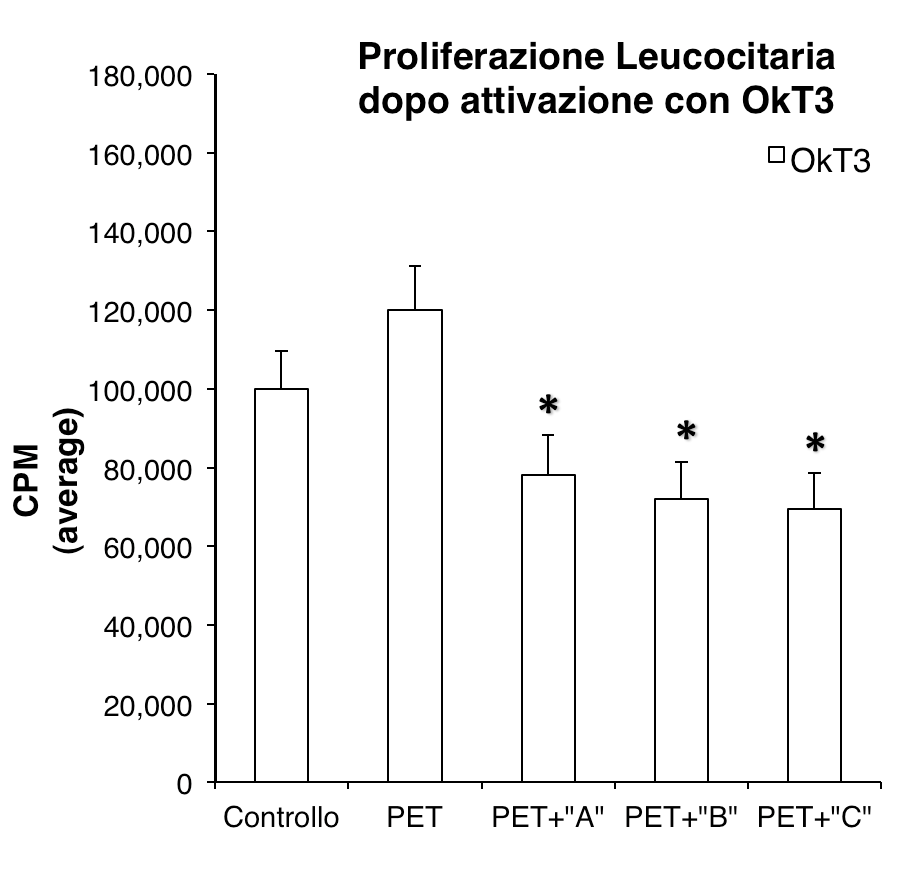
3) to measure the interference on the leukocyte proliferation triggered by exposure to a monoclonal antibody capable of activating the lymphocytes through interaction with the CD3 components of the TCR receptor complex.
4) to measure the ability of the surface treatment Ag / PEO-Like to induce leukocyte activation.
5) to measure the interference on the leukocyte activation induced on exposure to a monoclonal antibody capable of activating the lymphocytes through interaction with the CD3 components of the TCR receptor complex.
All tests were carried out using a polyethylene terephthalate (PET) prism inserted inside the polystyrene culture plates in which the cells were cultivated throughout the experiment period. Prisms with a square base of 1 mm thickness have been used with a surface of 1 cm2 (1 cm of side). The cell culture capsules were of the "multiwell" model with 24 circular wells with a diameter of 15 mm each able to accommodate a single prism per well that occupies 50% of the surface.
The PET prisms were subjected to three different surface treatments with Ag / PEO-Like. The treatments were performed on only one of the two basic prism faces. The treated faces were turned upwards when the prisms were placed in the wells to offer the maximum surface treated in direct contact with the cells.
The different treatments, indicated below with the letters A, B and C, differ for the following parameters:
Type "A" - double film of nanometric thickness: first film of Ag / PEO-Like (Ag: 5%) in turn covered with a barrier film of PEO-Like (character 70% PEO).
Type "B" - double film of nanometric thickness: first film of Ag / PEO-Like (Ag: 5%) in turn covered with a barrier film of PEO-Like (character 30% PEO).
Type "C" - single film, of nanometric thickness, of Ag / PEO-Like (Ag: 5%)
For each type of surface treatment, 30 prisms were made which were used to perform 3 independent triplicate tests. As a control, the same number of PET prisms were used without any surface treatment.
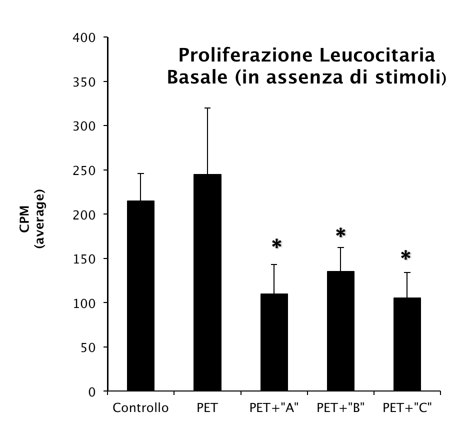
Test results:
Leukocyte proliferation test. The proliferation of PBMCs was assessed by incorporation of tritiated thymidine. Thymidine exposure for 6 hours occurred 48 hours after culturing and 6 hours before the dosage was performed.